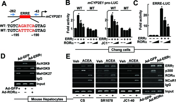
Figure 2.

CYP2E1 expression is controlled by RORα through an ERRE in the CYP2E1 promoter. (A) Schematic representation of the mouse CYP2E1 promoter with the wild-type (WT) or the mutant (MT) ERRE. (B) Chang liver cells were transfected with the WT or the MT CYP2E1 promoter-Luc reporter with empty vector, FLAG-ERRγ or FLAG-RORα for 24 h. Or the reporter transfected cells were treated with 10 or 20 μM JC1–40 (JC1) for additional 24 h. (C) Chang liver cells were transfected with the ERRE-Luc reporter with EV, FLAG-ERRγ or FLAG-RORα for 24 h. Luciferase activity that normalized to the corresponding β-galactosidase activity was converted to fold activity with no treatment as 1. The data represent mean ± SD. *P < 0.05 versus EV; #P < 0.05 versus ERRγ (n = 3). (D) The hepatocytes were infected by Ad-GFP or Ad-ERRγ with Ad-RORα for 24 h. (E) The primary cultures of mouse hepatocytes were treated with 20 μM ACEA, or infected by adenovirus encoding GFP or ERRγ. The hepatocytes were treated with 20 μM CS, 5 μM SR1078 or 20 μM JC1–40, or co-infected by Ad-RORα for 24 h as indicated. DNA fragments that contain flanking region of the ERRE on the CYP2E1 promoter were immunoprecipitated with indicated anti-histone antibodies (panel D), anti-ERRγ, anti-p300, anti-RORα or anti-NCoR1 antibodies (panel E) and then amplified by PCR using primers shown in panel A. Representatives of at least three independent experiments with similar results are shown.
