Figure 1.
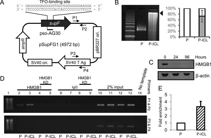
HMGB1 binds TFO-directed ICLs in human cells. (A) Schematic representation of the pSupFG1 plasmid containing the TFO pAG30-binding site within the supF gene. P1 and P2 indicate the locations of the forward and reverse primers proximal to the ICL; P3 and P4 indicate the locations of the control distal primer set. (B) Left panel, plasmid pSupFG1 (P) alone or pSupFG1 incubated with pAG30 and then irradiated with UVA at 1.8 J/cm2 (P-ICL) were resolved by 1% alkaline agarose gel electrophoresis to determine the efficiency of ICL formation (indicated by the black arrow). Right panel, quantification of three gels indicated that an average of ∼70% of the plasmid substrates contained TFO-directed ICLs. Error bars represent ± standard deviation (SD). (C) Western blot demonstrating the efficiency of HMGB1 depletion at 24 and 96 h (4 days) after siRNA treatment in U2OS cells. (D) PCR analysis of ChIP assays from U2OS cells demonstrated an enrichment of HMGB1 at the TFO-directed psoralen ICL region with proximal primers P1 and P2 (top panel) and distal control primers P3 and P4 (bottom panel). Lane 1, 100 bp ladder; lane 2, control plasmid (P); lane 3, ICL-containing plasmid (P-ICL). Lanes 4 and 5 are similar to lanes 2 and 3, respectively, but following siRNA-mediated depletion of HMGB1 from U2OS cells. Lanes 6, 7, 8 and 9 are IgG controls for lanes 2, 3, 4 and 5, respectively. Lanes 10–13 represent 2% input, lane 14 is a no template PCR control. (E) Quantification of PCR amplification from C indicated greater than three-fold enrichment of HMGB1 on the ICL-containing plasmid (P-ICL) compared to the control plasmid (P), n = 4, error bar represents ± SD.

