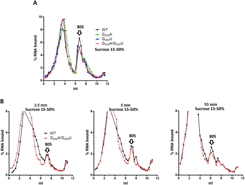
Figure 3.
80S initiation complexes assembly on mutant and WT IRES. 2 pmol of WT (black) or G266A (blue) or G268U (green) or DM (G266A/G268U, red) 32P- labeled RNAs were used to program 50 μl of RRL and 80S complexes were resolved by fractionation onto 15–50% sucrose density gradients. RRL were treated with cycloheximide before to observe 80S accumulation (A) The WT and double mutant RNAs were incubated with untreated reticulocyte lysate and translation was stopped after 2.5; 5 or 10 min addition of cycloheximide (B) 0 ml is the top of the gradient (lighter fraction) while 12 ml is the bottom (heavier fraction). The results are representative of at least three independent experiments. Arrows indicating the 80S complex were positioned according to control experiments carried out with a cap-dependent gene and according to UV profile of gradients carried out in the same conditions with purified 40S, 60S and 80S complexes (Supplementary Figure S7). The small higher density peak corresponds to a disome due to some leakage in the cycloheximide blockage.

