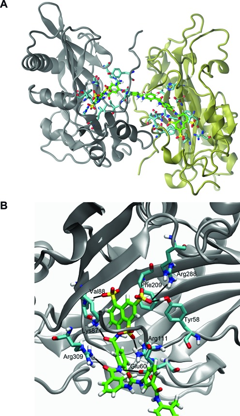
Figure 6.

Proposed binding mode of suramin to REL1. (A) Predicted binding of suramin to two REL1 enzymes simultaneously. The two enzymes have been colored gray and tan, respectively. Suramin is shown with its carbon atoms colored green. Only side-chains of residues in the two enzymes that interact with suramin are shown for clarity (carbon atoms colored cyan). (B) Detailed view of the predicted interaction of one half of suramin with key residues in the ATP binding pocket. Suramin, shown with carbon atoms colored green, assumes the binding position of REL1's natural substrate ATP and its interactions with the enzyme. Only the side-chains of interacting residues in the enzyme are shown for clarity (in cyan).
