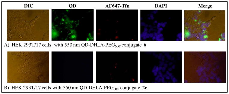
Figure 5.
Representative images of HEK 293T/17 cells incubated at 37°C for 1 hour with 50 nM of 550 nm QD DHLA-PEG600 conjugated to CPP-ligate 6 (A) or starter peptide 2c (B) as a control. For each panel the corresponding differential interference contrast (DIC), QD fluorescence (~550 nm), AF647-Tfn fluorescence (~670 nm), DAPI fluorescence (~460 nm), and merged composite images are shown.

