. 2015 Nov-Dec;41(6):1116–1125. doi: 10.1590/S1677-5538.IBJU.2014.0585

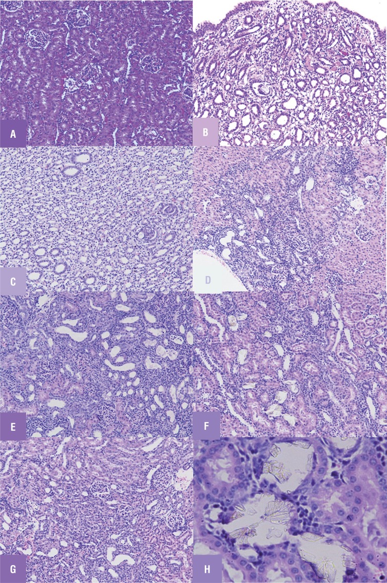
Figure 1. Light microscopic architecture of kidney showing (A) Renal tissue of control (group 1) rats showing no sign of crystal deposition. (B) Renal tissue of urolithiatic rats (group 2) showing crystals deposition and severe obstructive nephropathy (C) Renal tissue of (group 3) STS treated rats showing mild crystal deposition with mild nephropathy. (D) Renal tissue of (group 4) diazoxide treated rats showing mild crystal deposition with moderate obstructive nephropathy (E) Renal tissue of (group 5) glibenclamide treated rats showing prominent crystal deposition with severe obstructive nephropathy (f) Renal tissue of (group 6) diazoxide +STS treated rats showing crystal deposition with severe obstructive nephropathy (g) Renal tissue of (group 7) glibenclamide + STS pretreated rats low crystal deposition with mild nephropathy (h) Crystal's deposition as observed under 40X zoom.

Figure 1