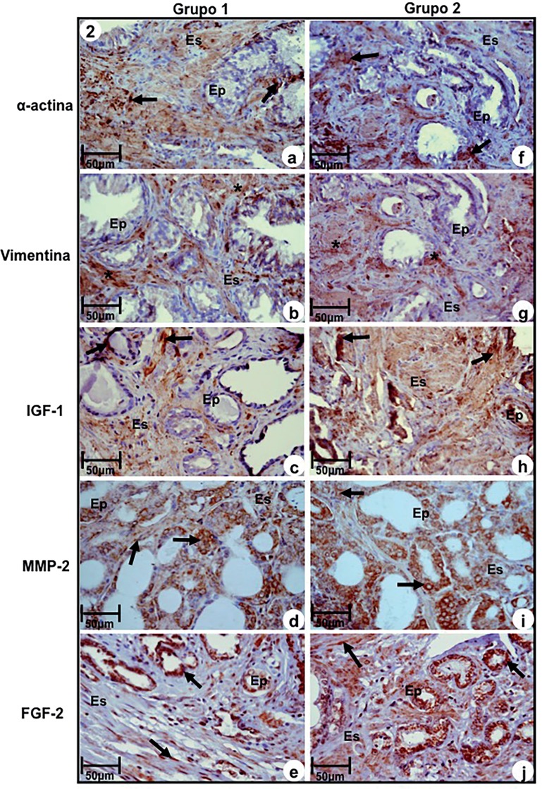
Figure 2. Immuno-staining of antigens α-actin, Vimentin, IGF-1, MMP-2 and FGF-2 at prostatic peripheral zone of Groups-1 (a, b, c, d, e) and 2 (f, g, h, i, j). (a) and (f) Immunoreactivity to α-actin (arrows). (b) and (g) Immunoreactivity to Vimentin (asterisks) in myofibroblasts. (c) and (h) Imunoreactivity to IGF-1 (arrows) in epithelium and stromal compartments. (d) and (i) Immunoreactivity to MMP-2 (arrows) in epithelial and stromal compartments. (e) and (j) Immunoreactivity to FGF-2 (arrows) in cells of secretory epithelium and fibroblasts of stromal compartment.
a-j, Ep–secretory epithelium; Es-stroma. Scale of 50μm.

