. 2015 Jul-Aug;41(4):773–790. doi: 10.1590/S1677-5538.IBJU.2013.0148

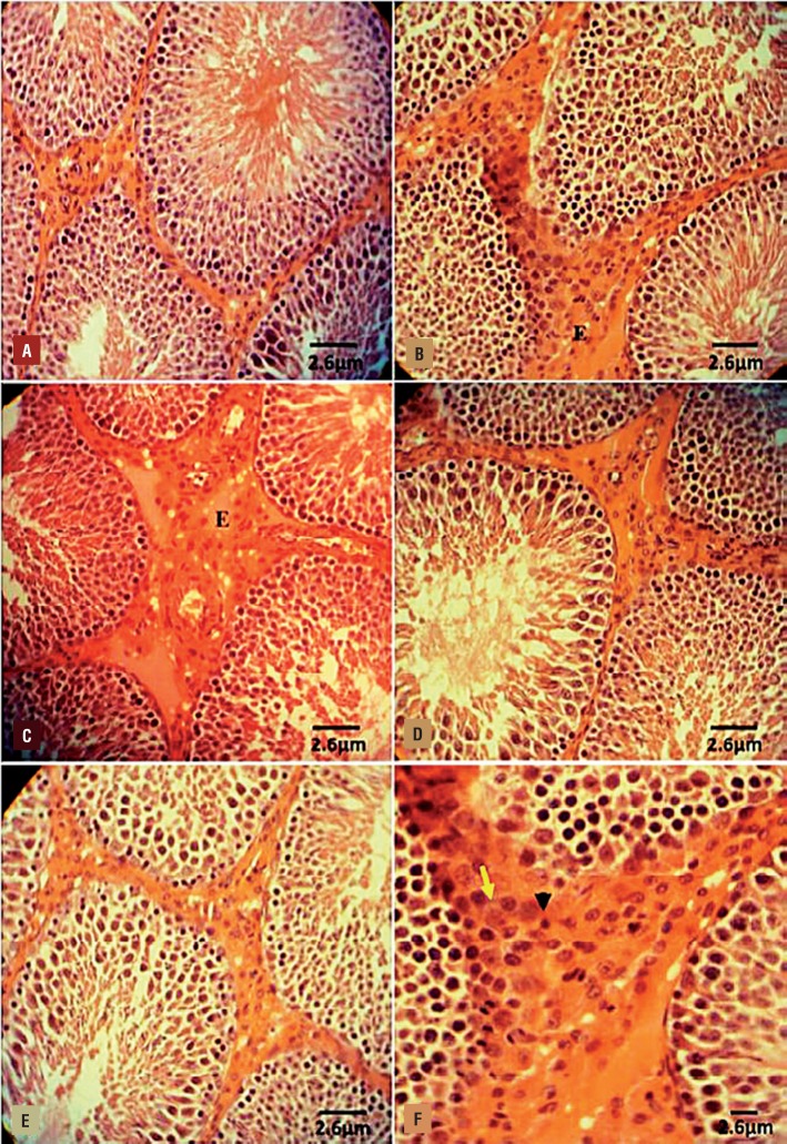
Figure 2. Photomicrograph of rat testes. (A) Control-sham group: normal germinal epithelium with no edematous connective tissue; (B) VCL-induced group: testicular tissue represented with severe edema (E) in connective tissue and remarkable immune cells infiltration in interstitial tissue; (C) VitE-administrated group: the protected seminiferous tubules with positive TDI and RI are observed, while the edematous interstitial tissue (E) is remained unchanged; (D) DEX received group: note decreased edema and down regulated immune cells infiltration. The seminiferous tubules are presented with normal TDI and RI; (E) VitE+DEX-administrated group: the edema and immune cells infiltration significantly reduced versus figures D and E, and the tubules are presented close to normal; and (F) Higher magnification from interstitial tissue of non-treated VCL-induced testis: note mononuclear immune cell (head arrow) infiltration in edematous tissue and hypertrophied leydig cells (arrow) distribution. Iron-Weigert staining technique (×400).

Figure 2