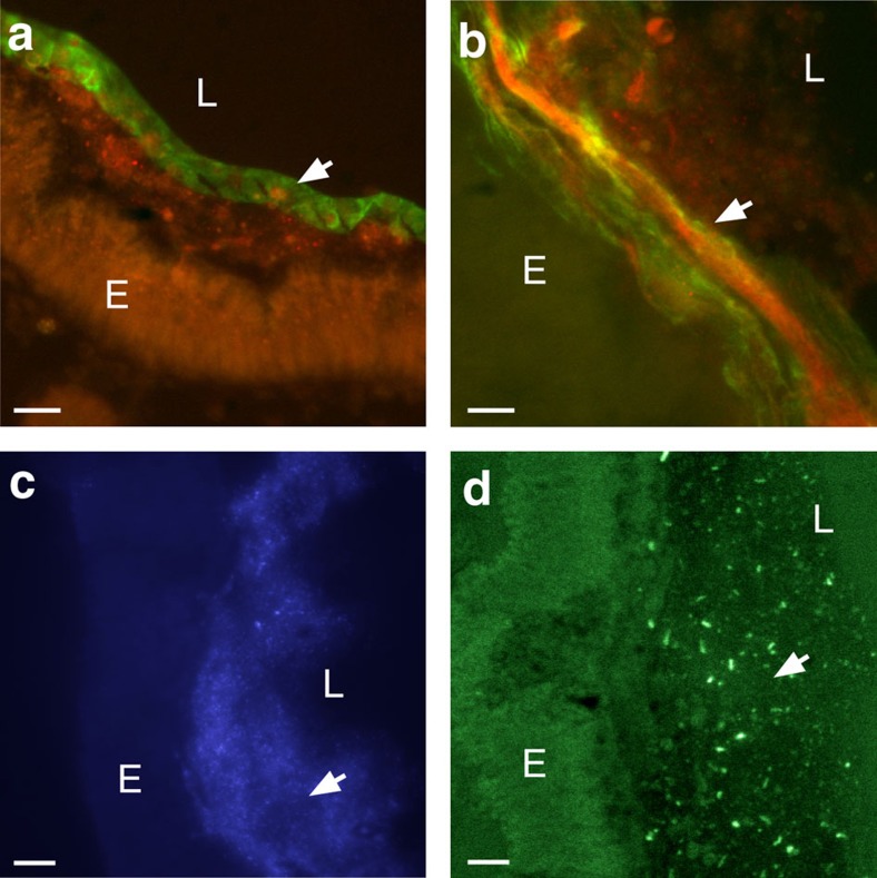
Figure 4. Detection of VCBP-C in both dense and loosely associated mucus.
(a,b) Colocalization (yellow-merged signal) of chitin (Fc-CBD-C DyLight 488, green) and VCBP-C (Alexa Fluor 594, red). Dense mucus (glycocalyx-like) of the midgut can form (a) ribbon-like structures (arrow) as opposed to (b) less dense mucus (arrow) seen in the distal gut. (c) Microbiota-sized particles (arrow) seen in the mucus detected by Hoechst staining of DNA were confirmed as bacteria by 16S FISH (arrow) (d). Staining is negative with isotype and secondary antibody controls. Scale bars,10 μm. E, epithelium; L, lumen.

