Fig. 2.
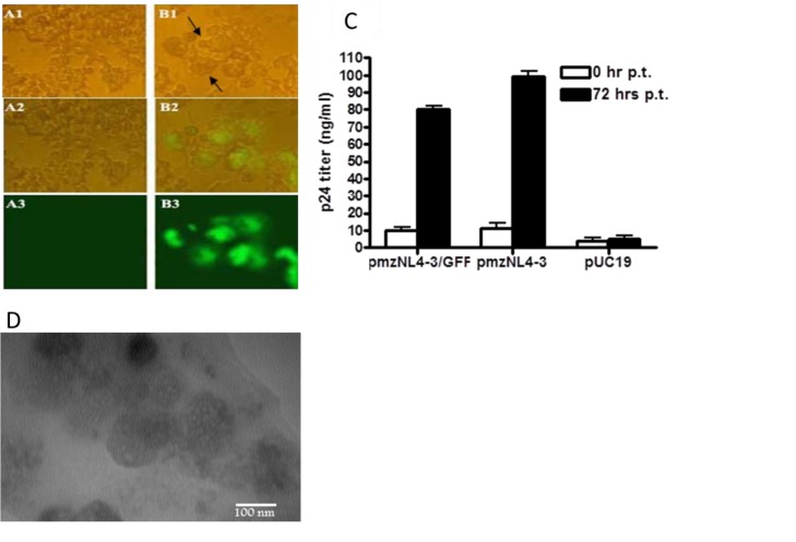
Production of the first generation GFP-mzNL4-3 virions. In contrast to the cells transfected with pUC19 control plasmid (A1-A3), cells co-transfected with pmzNL4-3/GFP and helper plasmids (pSPAX2, pMD2G) produced large round syncytia (B1, shown with arrows) and GFP-emitting (B1-B3) cells at 72 hrs post-transfection, which were observed under the immunefluorescence microscope (400x). p24 ELISA assay of culture supernatant at 72 hrs post-transfection (p.t.) indicated the secretion of p24 for pmzNL-43/GFP-transfected cells comparable to the positive control cells transfected with pmzNL4-3 plasmid. Again cells receiving pUC19 produced a background level of p24. Bars show mean of p24 concentration ± SD in three transfected wells (C). Electron microscopy also confirmed the formation of 120-150 nm particles within the culture supernatant of pmzNL4-3/GFP cells (D).

