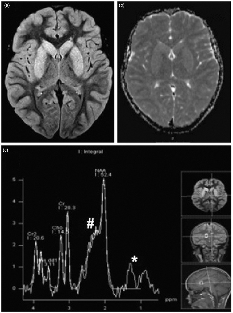
Figure 1.
(a) Axial FLAIR image shows hyperintense signal of the basal nuclei, bilateral and symmetric. (b) ADC map demonstrates diffusion facilitation in the basal nuclei. (c) Proton MRS from right caudate nuclei at TE of 30 ms reveals the presence of lactate/lipid (*) and elevation in excitatory neurotransmitters (#). The case originally appeared on the AJNR website.

