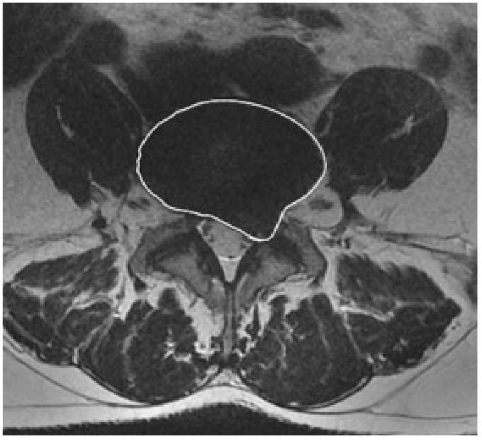
Figure 3.
Axial MR image of the patient described in Figure 1. FSE T2-weighted MR image shows entire disc and herniated parts included in the ROI for IDVA.
(MR: magnetic resonance; FSE: fast-spin echo; ROI: region of interest; IDVA: intervertebral disc volumetric analysis.)

