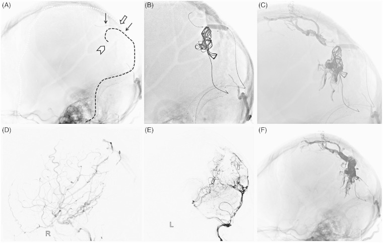
Figure 3.
First stage operation. A) Unsubtracted lateral view showed the Hyperglide balloon position (black arrow), and the enchelon-10 microcatheter navigated into the venous pouch on the falx (catheter tip, arrowhead; catheter, black dotted line). A Marathon microcatheter catheterized the distal middle meningeal artery (catheter tip, white arrow; catheter, white dotted line). B) The balloon was inflated with the distal one-third in the venous pouch and the proximal two-thirds in the SSS, and several coils were employed through the Echelon-10 microcatheter. C) Onyx was injected through the Marathon microcatheter and Echelon-10 microcatheter alternately. Final right CCA lateral angiography (D) and left VA lateral angiography (E) showed partial obliteration of the fistula and blood flow slowed down. F) Post-embolization cast was seen on unsubtracted lateral view.

