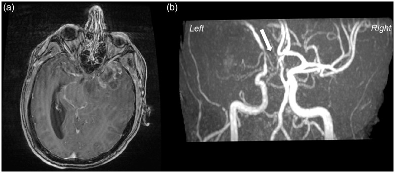
Figure 2.
T1-weighted MRI, post-gadolinium (a) demonstrates a large necrotic lesion in the left frontotemporal region, typically ring-like, enhanced with perilesional edema, suggesting of malignant brain tumor. (b) MR angiography by TOF reveals the uprising and partial stenosis of the left middle cerebral artery (MCA) by a tumor and the major stenosis of A1 segment of anterior cerebral artery (ACA) (white arrow).

