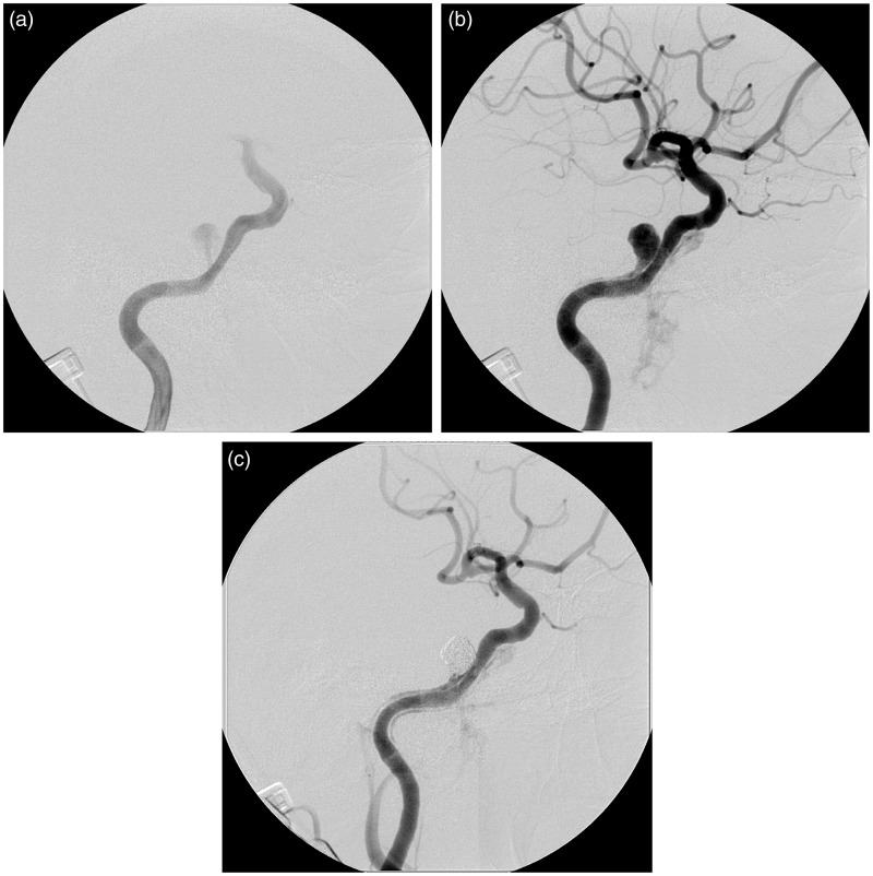
Figure 3.
Right internal carotid angiography. (a) Early arterial image demonstrating the shunt from the ICA into the newly developed aneurysm. (b) Late arterial phase demonstrating the retrograde blood flow in the pseudolumen along the internal carotid artery, suggestive of the long dissection of the internal carotid artery. The amount of the shunt flow into the cavernous sinus and the pterygoid plexus is much smaller compared with that recognized in the diagnostic angiography. (c) The final image revealing complete occlusion of the aneurysm with minimal shunt flow.

