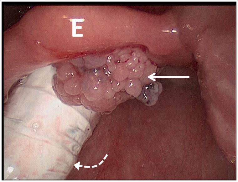
Figure 1.
Intra-operative photograph from laryngoscopy reveals an approximately 1 cm exophytic, pedunculated, papillomatous lesion (arrow) on the laryngeal surface of the suprahyoid epiglottis (E), close to midline, without apparent involvement of the underlying cartilage. The endotracheal tube is seen posteriorly (curved dotted arrow).

