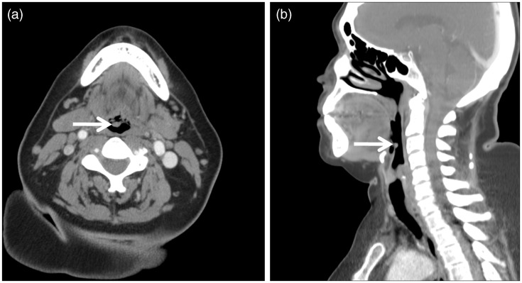
Figure 2.
Axial (a) and sagittal (b) contrast-enhanced computed tomography (CT) images of our 65-year-old female patient revealed a 0.7 cm × 0.5 cm × 0.5 cm (transverse × anterior-posterior × craniocaudal) circumscribed structure (arrow) of approximately 50 Hounsfield units, inseparable from the posterior (laryngeal) surface of the right parasagittal epiglottis. The epiglottis was not thickened, and the remainder of the larynx appeared normal.

