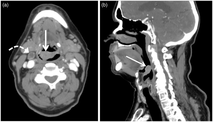
Figure 4.
Axial (a) and sagittal (b) contrast-enhanced computed tomography (CT) images of a 73-year-old male patient with foreign body sensation revealed a 2.6 cm × 1.5 cm × 2.0 cm (transverse × anterior-posterior × craniocaudal) epiglottic mass (arrow) of approximately 70 Hounsfield units biopsy proven squamous cell carcinoma. A necrotic lymph node was also identified (curved arrow).

