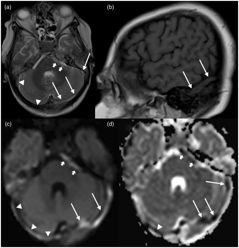
Figure 1.
MRI of a 10-year-old girl with extensive CSVT: (a) axial T2-weighted image shows hyperintense signal in the left (long arrows) and right (arrowheads) transverse and sigmoid sinus and mixed hyper- and isointense signal in the left cavernous sinus (short arrows); (b) sagittal T1-weighted image reveals isointense signal in the left transverse sinus (long arrows); (c) axial trace of diffusion and (d) apparent diffusion coefficient (ADC) maps show DWI-bright signal in the left (long arrows) and right (arrowheads) transverse and sigmoid sinus and the left cavernous sinus (short arrows) with matching low ADC values.

