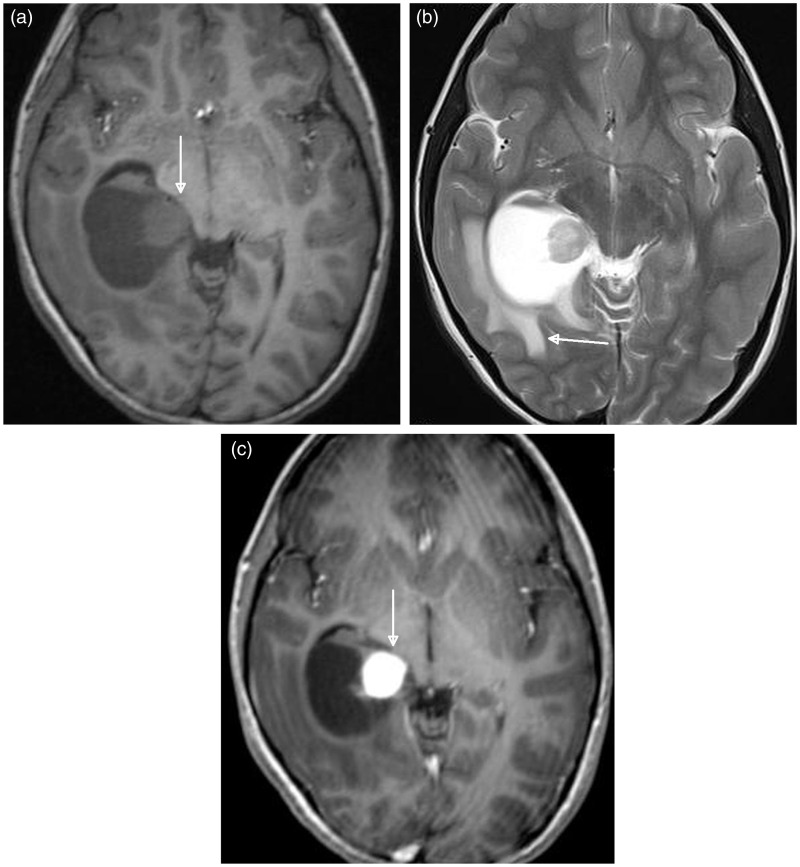
Figure 1.
Axial plane brain MRI findings of a 6-year-old female with desmoplastic non-infantile ganglioglioma. (a) T1-weighted image shows a large mass lesion in the right parahippocampal region adjacent to the ambient cistern. The cystic mass lesion has a mural nodule medially (arrow). MRI: magnetic resonance imaging. (b) T2-weighted image shows that the mural nodule is slightly more hyperintense than the grey matter. Note the transependymal flow of cerebrospinal fluid to the surrounding periventricular white matter (arrow). (c) Contrast-enhanced T1-weighted image demonstrates avid enhancement of the mural nodule (arrow).

