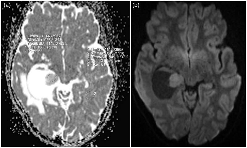
Figure 2.
(a) The nodule shows high ADC values compared with those of the white matter of the contralateral side. (b) Axial plane DWI of the brain demonstrates a slightly hyperintense signal of the mural nodule compatible with T2 shine affect. ADC: apparent diffusion coefficient; DWI: diffusion-weighted imaging.

