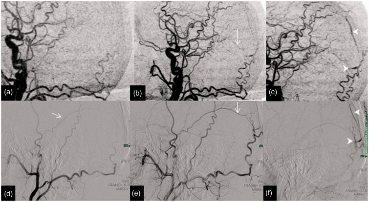
Figure 4.
Imaging of a 65-year-old man with an occipital haemorrhage; 4D-CTA and DSA illustrate the dural arteriovenous fistula of the falx. The upper row of images (a), (b) and (c) of lateral MIP of 4D-CTA shows the middle meningeal artery (arrow) supply to the fistula and the early draining occipital cortical vien (arrow head). The lower row of images (d, e and f) of the DSA in lateral projection display similar findings to 4D-CTA. This is an example of a Lariboisiere type III arteriovenous fistula, with direct cortical drainage. 4D-CTA: four-dimensional computed tomography angiography; MIP: maximum intensity projection; DSA: digital subtraction angiography.

