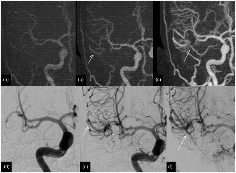
Figure 5.
Imaging of a 54-year-old man with a right temporal haemorrhage in which 4D-CTA and DSA highlight the AVM. The upper row of images (a, b and c) of coronal MIP of 4D-CTA demonstrates the early draining superficial middle cerebral vein (arrows) from the AVM. The lower row of images (d, e and f) of the DSA in anteroposterior projection display similar findings to the 4D-CTA. This is an example of a Spetzler–Martin grade 1 AVM with a nidus size of less than 3 cm with a superficial venous drainage. AVM: arteriovenous malformation; 4D-CTA: four-dimensional computed tomography angiography; MIP: maximum intensity projection; DSA: digital subtraction angiography.

