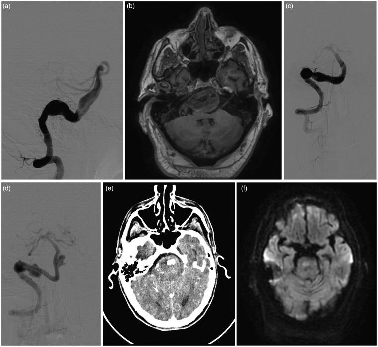
Figure 1.
(a) Lateral view of the initial angiogram showing a right VB junction aneurysm extending to the distal third of the basilar artery. (b) MRI T1 weighted image showing a fusiform aneurysm with compression of the brainstem. (c) A 3 month follow-up angiogram shows a partially thrombosed fusiform aneurysm. (d) A 10 month follow-up angiogram with development of endoleaks at the proximal and distal ends with growth of the aneurysm. (e) A 10 month CT angiogram shows hypervascularity at the periphery of the aneurysm that was not present on the initial CT angiogram, suggestive of blood flow to the vasa vasorum. This could be responsible for growth of the aneurysm. (f) Diffusion-weighted image showing ischemic changes in the brainstem.

