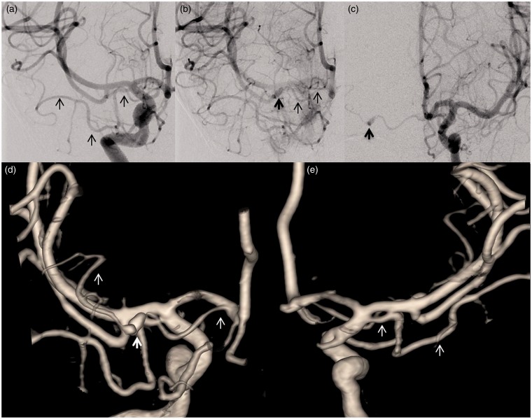
Figure 2.
Cerebral angiography demonstrating aMCA and dissecting aneurysm in type-3 aMCA. (a) Right ICA injection showing type-1 aMCA with cortical branches. (b) Late filing of dissecting aneurysm (bold dark arrow) of type-3 aMCA. (c) Early brisk filling of dissecting aneurysm of type-3 aMCA from a contralateral injection. (d) Three-dimensional (3D) surface shaded display (SSD) demonstrating right type-3 aMCA harboring a dissecting aneurysm (bold white arrow). (e) 3D SSD right type-1 aMCA (posterior view).

