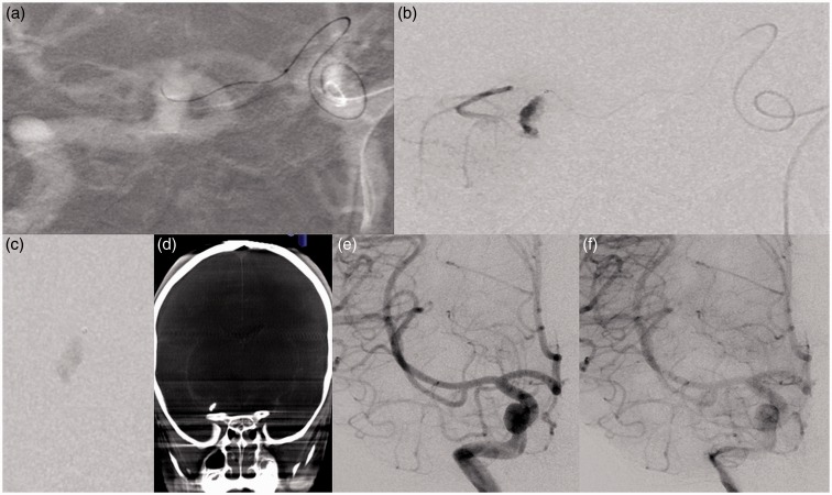
Figure 3.
Technical aspects and post procedure cerebral angiography. (a) Catheterization of type-3 aMCA from contralateral side using Magic 1.2 FM microcatheter. (b) Microcatheter injection showing filling of dissecting aneurysm and distal cortical territory. (c) Glue cast seen in the aneurysm sac. (d) Dyna CT showing glue cast within the aneurysm sac. (e, f) Early and delayed run showing obliteration of the aneurysm sac.

