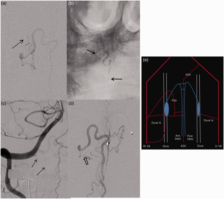
Figure 2.
Angiographic findings of complex arteriovenous fistula (AVF) at craniocervical junction. (a) Superselective angiography reveals a right-sided AVF that drained mainly into the anterior medullary vein (the black dotted arrow indicates the distal tip of the microcatheter). (b) Transarterial embolization using histoacryl glue was conducted (the black arrows refer to the proximal and distal points of the histoacryl glue). (c) A post-embolization vertebral angiogram reveals that the remaining AVF was fed by the dural branches (black arrow) of the vertebral artery (VA). (d) Superselective anterior spinal angiography revealed that the right AVF was supplied by branches of the ASA. The distal portion of the ASA branch has a common channel with the initial embolized feeding artery (The black hollow arrow indicates the common channel). (e) A schematic diagram illustrating both AVFs. (A, artery; ASA, anterior spinal artery; PSA, posterior spinal artery; PMV, perimedullary vein; VA, vertebral artery).

