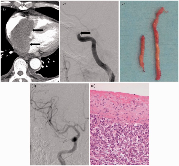
Figure 1.
A 55-year-old man with a cardiac tumor and hyperacute stroke of right middle cerebral artery territory.
(a) Contrast-enhanced chest CT scan shows a well-defined solid tumor in the left atrium (arrows).
(b) Cerebral angiography shows a complete occlusion of the right distal internal carotid artery (arrow).
(c) Gross appearance of emboli shows yellowish, gelatinous, and hard tissue.
(d) Final angiogram after aspiration thrombectomy using Penumbra catheter demonstrating complete recanalization of the right internal carotid artery and middle cerebral artery territories.
(e) Histologic section showing high-grade undifferentiated sarcoma (HE, x 400).

