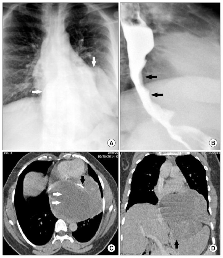
Fig. 1.
(A) Chest radiograph showing a well-defined retrocardiac soft tissue mass (white arrows) with intra-abdominal extension. (B) Barium swallow showing smooth luminal narrowing of the distal esophagus and gastroesophageal junction due to extrinsic compression by the mass (black arrows). (C) Axial contrast-enhanced CT image showing a large lobulated hypodense well-defined mass (white arrows) containing areas of calcification (black arrow). (D) Coronal reformat of CT scan showing the mass extending through the esophageal hiatus and compressing the fundus of the stomach (black arrow). CT, computed tomography.

