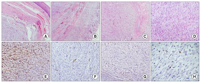
Fig. 2.
Photomicrographs show (A) a well-circumscribed spindle cell lesion arising from the lower end of the esophagus (H&E, ×40) and (B) reaching up to the gastric fundus (H&E, ×40). (C) The mass is lobulated (H&E, ×40) and (D) shows fascicles with spindle cells showing hyperchromasia and nuclear buckling on a myxoid background (H&E, ×100). On immunohistochemistry, (E) these cells were positive for S100 protein (S100, ×40) and (F, G) negative for CD34 and DOG 1 stains (CD34 and DOG-1, ×100). (H) The Ki67 labeling index is low (Ki67, ×100).

