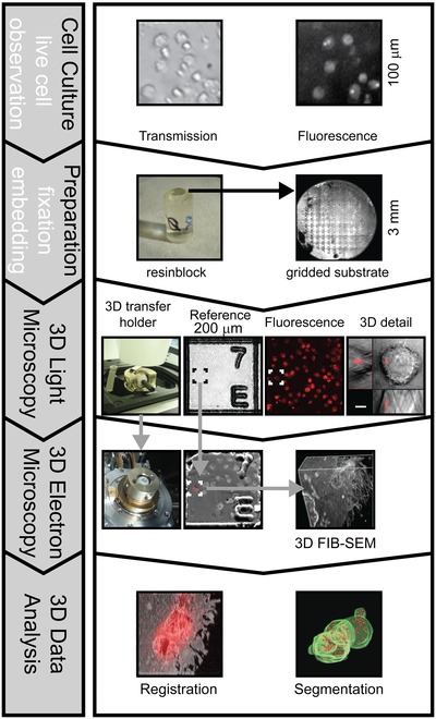
Figure 1.

Generalized scheme of the correlative 3D workflow.
Since fluorescence is preserved during the EM preparation process, 3D confocal fluorescence microscopy allows selection and detailed imaging of target structures within the resin block. Additional recording of coordinate marks on the block surface allows the transfer of target coordinates to guide FIB‐SEM imaging with micrometre precision. Defined orientation of the block surface is ensured by the 3D transfer holder, which allows to align the block surface perpendicular to the z‐axis of the light microscope and to transfer directly the sample to the electron microscope. Exact guidance of FIB‐SEM by 3D light microscopy data restricts FIB‐milling and SEM imaging to target structures, minimizing processing time and data collection. Finally this simplifies the task to register light and electron microscopic datasets.
