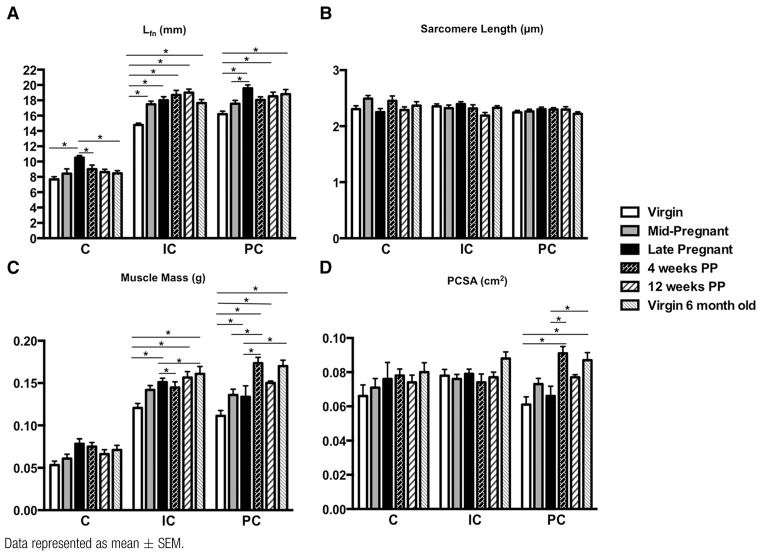
FIGURE 1.
Comparison of architectural parameters of C, IC, and PC among virgin, pregnant, and PP groups
Data represented as mean ± SEM.
C, coccygeus; IC, iliocaudalis; Lfn, fiber length normalized to optimal sarcomere length; PC, pubocaudalis; PCSA, physiologic cross-sectional area; PP, postpartum.
*Significantly different P values derived from 2-way analysis of variance, followed by Tukey pairwise comparisons with significance level set to 5%.
Alperin. Pregnancy adaptations in rat pelvic muscles. Am J Obstet Gynecol 2015.

