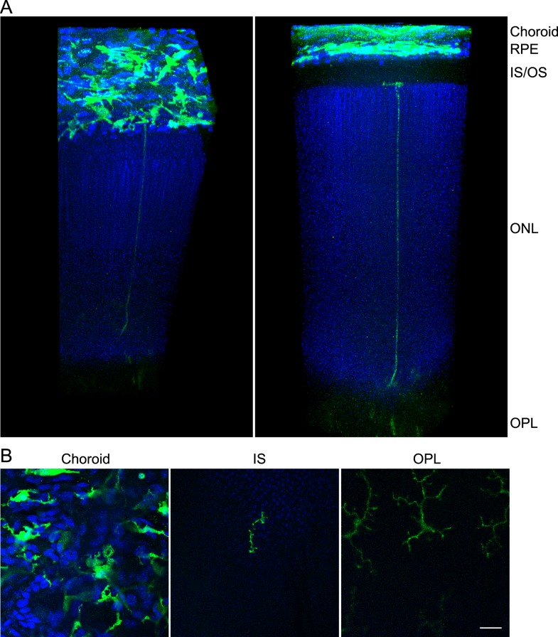
Figure 7.
GFP cells extending from sclerochoroid to OPL in retina of CX3CR1+/GFP mouse. (A) Confocal images from sclerochoroid to OPL of retina were reconstructed in a 3D view using Volocity software. RPE/retina whole mounts of 2-month-old CX3CR1+/GFP mice were immunostained with GFP antibody after 10% H2O2 pretreatment at 55°C for 1.5 hours or 4°C for 7 days. These representative images were taken from 55°C H2O2 pretreatment. Left is the tilted view of right panel to better display the horizontal plane of the sclerochoroid. (B) Original confocal images. From a total of 1190 z stack images, the 58th, 138th, and 993rd single images were taken for the presentation of CX3CR1+/GFP macrophage/microglia in choroid, IS, and OPL. IS/OS, inner and outer segments of photoreceptors. Scale bar: 20 μm.

