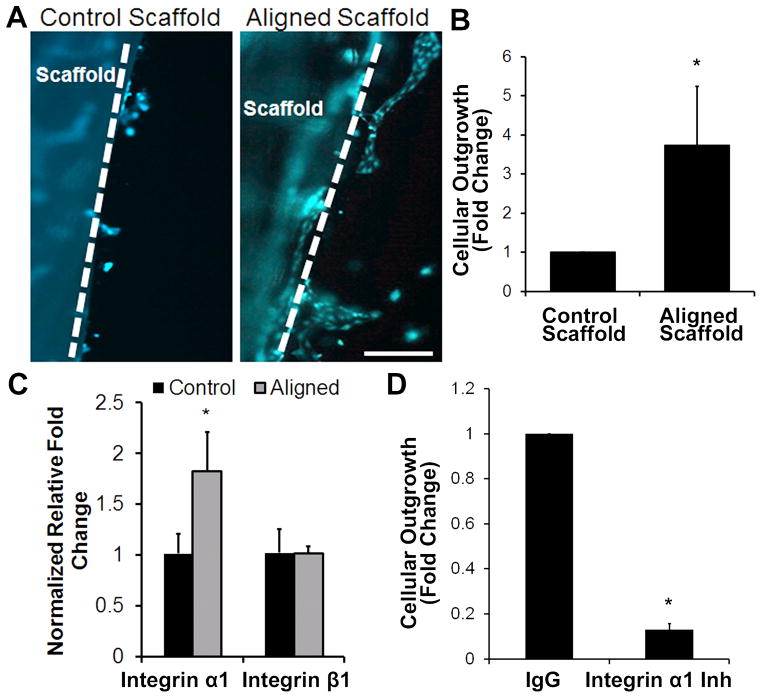
Fig. 3. Endothelial outgrowth from aligned nanofibrillar scaffolds.
Human ECs seeded on fibronectin-pre-coated control or aligned scaffold were encapsulated into a 3D hydrogel for tracking cellular outgrowth. (A) Fluorescently labeled ECs are shown migrating from scaffold into the surrounding hydrogel after 3 days. Dotted line denotes border of scaffold. (B) Quantification of cellular outgrowth from control or aligned scaffold after 3 days (n=3, *P<0.01). (C) qPCR analysis of integrin subunit gene expression (n=3, *P<0.05). (D) Cellular outgrowth from aligned nanofibrillar scaffolds in the presence of integrin α1 inhibition antibody or IgG control (n=3, *P<0.001). Scale bar: 200 μm.

