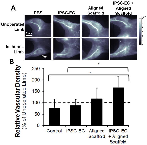
Fig. 5. Near infrared-II (NIR-II)-based fluorescence imaging of the hind limb vasculature at 28 days after implantation of aligned scaffold with human iPSC-ECs.
(A) Representative images of each group for the ischemic and unoperated limb. (B) Quantification of relative vascular density between treatment groups. Dotted lined denotes vascular density of unoperated limb (*P<0.05, n≥3).

