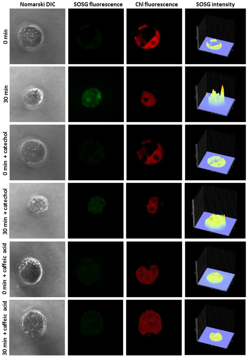
Figure 6. Detection of singlet oxygen in Chlamydomonas cells by laser confocal scanning microscopy.
The formation of 1O2 was measured in non-heated and heated Chlamydomonas cells in the presence and the absence of catechol and caffeic acid using fluorescent probe SOSG. Heated cells were treated for 30 min in a water bath at 40 °C under dark. The images represent from left to right: Nomarski DIC, SOSG fluorescence, chlorophyll fluorescence and integral distribution of SOSG signal intensity (0–4096) in the 12-bit microphotographs.

