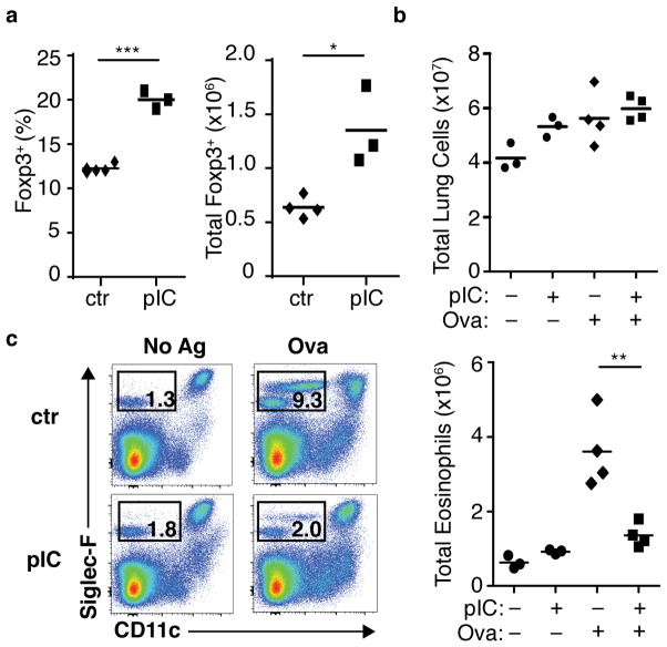
Figure 1. Non-specific bystander inflammation results in increased Foxp3+ Treg cells and suppression of primary antigen-specific mucosal inflammatory response.
(a) Wild-type (WT) BALB/c mice were treated with poly(I:C) (pIC) or PBS (ctr) intranasally for two consecutive days. Seven days after the first treatment, frequency (left panel) and total number (right) of Foxp3+ CD4+ T cells were assessed. Each symbol represents an individual mouse, data are representative of two independent experiments. (b–c) Primary antigen-specific pulmonary inflammation following intranasal poly(I:C) (described in Materials and Methods and Supplementary Fig. 1f). (b) Total pulmonary cell counts for mononuclear cells. (c) Pulmonary eosinophil infiltration, representative flow cytometry plots from indicated mice (left), total eosinophil counts for indicated mice (right). Each symbol represents an individual mouse, data representative of two independent experiments, n ≥ 6. Mice pre-treated with pIC (+) or PBS (−) and challenged with OVA–TSLP (+) or PBS (−) as indicated below the axis. P-values by student’s two-tailed t-test, * P <0.05, ** P <0.01, *** P <0.001.

