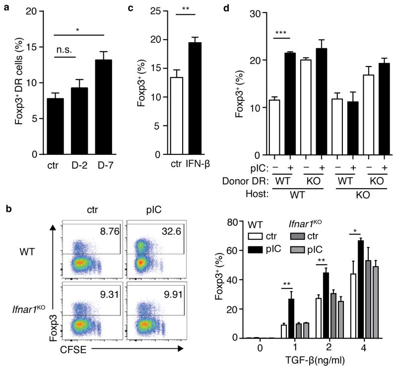
Figure 5. Antiviral bystander inflammation and type I interferon conditions naïve T cells for enhanced de novo Treg cell differentiation.
(a) DR cells were parked in BALB/c host mice, which were then inoculated with LCMV at seven days (D-7) or two days (D-2) prior to harvest; control mice (ctr) were left untreated. CD4+ T cells were isolated and cultured with OVA-pulsed APC and TGFβ for 5 days. Bar graphs indicate %Foxp3+ among cultured donor DR cells. (b) CD4+ T cells from control (ctr) and poly(I:C)-treated (pIC) wild-type (wt) and IFNaRKO mice were prepared as in Supplemental Fig.3a. After 5 days, cells were stained for Foxp3. Left panels, representative FACS plots; right, bar graph Foxp3+ DR frequencies. (c) DR splenocytes were incubated for 48 hours with PBS (ctr) or rIFN γ (IFN γ), then CD4+ T cells were purified and incubated with OVA-pulsed APC and TGF β. After 3 days, DR cells were assessed for Foxp3. (d) WT or IFNaRKO DR cells were transferred into WT or IFNaRKO Balb/c hosts, which were then treated with PBS (ctr) or poly(I:C) as indicated. Lymphocytes were cultured with Ova-pulsed APCs and TGF β for 5 days, then assessed for intracellular Foxp3. Bar graphs depict mean plus SEM of triplicate values, data are representative of two independent experiments, n ≥ 2 mice per group. P-values by student’s two-tailed t-test, *p<0.05, **p<0.01, ***p<0.001.

