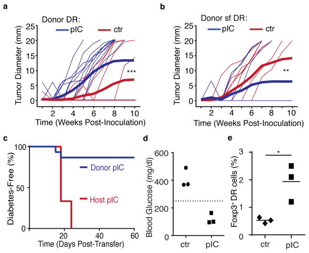
Figure 6. Bystander inflammation conditioning of naïve CD4+ T cells inhibits antigen-specific CD4+ T cell responses in a Foxp3-dependent manner.
(a) CD4+ T cells from control (ctr) or poly(I:C)-treated (pIC) DR mice were transferred into Balb/c hosts. Mice were immunized with OVA peptide and 7–10 days later were inoculated subcutaneously with A20-tGO lymphoma cells, and tumor growth was monitored. Thin lines represent measurement from individual mice, thick lines represent cumulative mean tumor size. (b) As in part (a), but donor cells were Foxp3-mutant scurfy (sf) DR CD4+ T cells. (c) RIP-mOva/RAGKO host mice received ICTN DR cells and were left untreated (Donor pIC), or received ctr DR cells, and host mice were injected with pIC (Host pIC), and blood glucose was monitored. Graph shows diabetes-free survival. (d–e) Ctr or ICTN DR CD4 T cells were transferred into RIP-mOva/RAGKO host mice, all mice were treated with pIC, and donor DR cells were assessed 20 days later (summarized in Supplementary Fig. 6d). Each symbol represents a single mouse, data from one of two independent experiments shown, n=6. (d) Blood glucose levels at day 20, dashed line indicates the threshold for clinical diagnosis of diabetes. (e) Frequency of Foxp3+ donor DR cells in the pancreatic lymph nodes at day 20. Data compiled from (a) three independent experiments, n=14 or (b,c) two independent experiments, n=10. P-values by (a,b) Two-way ANOVA or (f) student’s two-tailed t-test, *p<0.05, **p<0.0005, ***p<0.0001.

