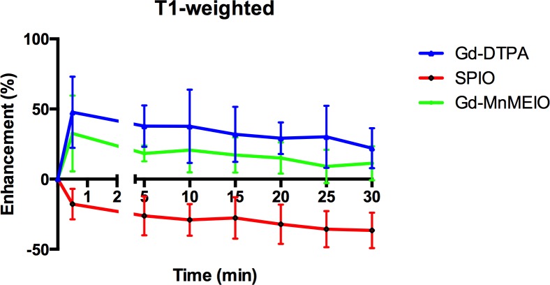
Fig 6. Time course curves of liver parenchyma enhancement on T1-weighted images by using Gd-DTPA (n = 8), SPIO (n = 8), and Gd-MnMEIO (n = 8).
The error bars indicate standard deviation. Contrast enhancement with Gd-DTPA and Gd-MnMEIO is significantly better than with SPIO at every post-enhancement time point (p <0.001), while no significant difference can be observed between Gd-DTPA and Gd-MnMEIO (p > 0.05).

