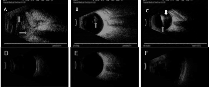
Fig 3. B-scan images of the patients' eyes infected with B. cereus before (A,B,C) and after (D,E,F) surgery operation.
(A). Infection caused by GTI strains showed severe vitreous opacity (gay arrows). (B). Infection caused by GTII strains showed mild vitreous opacity (gray arrows). (C). Mild vitreous opacity infection caused by GTIII strains (gray arrow),high reflection and ascoustic shadow in vitreous showed foreign body (white arrow). (D). Infection caused by GTI strains after surgery. (E). Infection caused by GTII strains after surgery. (F). Infection caused by GTIII strains after surgery(the false expansion of eye ball after vitreous surgery with silicon oil tamponade).

