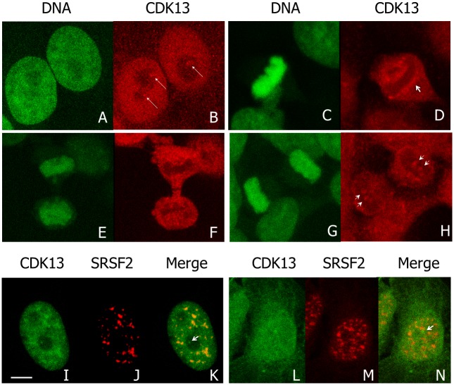
Fig 1. Distribution of CDK13 during cell cycle.
Confocal images of HeLa (A-H) cells in which DNA is stained with SYBR green (A,C,E,G) and CDK13 was detected by immunolabeling using anti-CDK13 antibody (B,D,F,H). In interphase (A,B), arrows highlights brighter spots of CDK13 close to the nucleoli. CDK13 distribution is further described in metaphase (C,D), early (E,F) and late telophase (G,H). During mitosis, arrowheads highlight accumulation of CDK13 close to the chromatin (D) and show CDK13 localized in dots in late telophase (H). A co-labelling of CDK13 and SRSF2 (I-N) shows the enrichment of CDK13 in speckles (SRSF2 positive spots) and in dots negative for SFSF2 (arrows in K,N) in the nucleolar area of HeLa (I-K) and U2OS (L-N) cells. Scale bar: 5μm.

