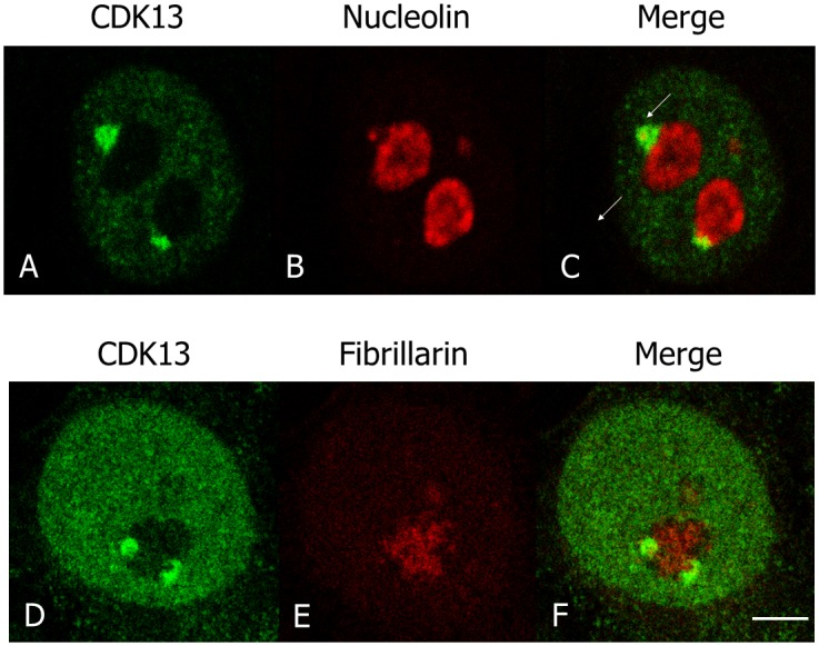
Fig 2. Localization of CDK13, nucleolin and fibrillarin during interphase.

In HeLa cells, the localization of CDK13 (A,C,D,F), nucleolin (B,C) and fibrillarin (E,F) were detected using rabbit anti-CDK13 and mouse anti-nucleolin or anti-fibrillarin antibodies. A poor (C) or absent (F) colocalization of CDK13 with these nucleolar markers demonstrates that CDK13 is located in a perinucleolar structure. Scale bar: 5μm.
