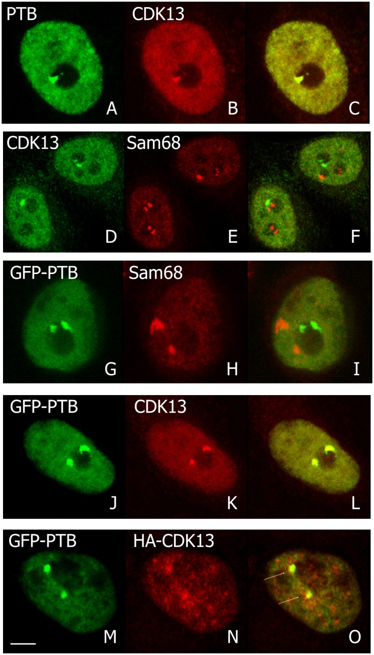
Fig 4. Colocalization of CDK13 with perinucleolar structures in interphase.

Endogenous CDK13 (B,D), PTB (A) and Sam68 (E) immunolocalizations in HeLa cells are shown in confocal optical sections and colocalization appeared in yellow in merge images (C,F). The CDK13 dots correspond to the PTB localization. Pictures G-I confirmed that PTB, expressed as a GFP-fusion protein, and Sam 68 are localized in different subnuclear domains. Overexpressed GFP-PTB (J,M) was co-localized with the endogenous (K) or overexpressed (N) CDK13 as visualized in the respective merge images (L,O). Arrows indicate the PNC in the merge optical sections (O). Scale bar: 5μm.
