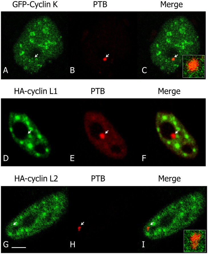
Fig 6. Localization of Cyclins K and L in interphase.

GFP-cyclin K and HA-cyclins L1 and L2 were expressed in HeLa cells and their localizations were analysed by fluorescence microscopy using respectively GFP-cyclin K (A) or immunofluorescence with anti-cyclin L1 (D) and L2 (G) antibodies. Localizations were compared with the one of endogenous PTB (B,E,H). Respective merge images (C,F,I) show an absence or a very poor co-localization of cyclins with CDK13 in PNC. An increased magnification of merge labelling is inserted in C and I. Scale bar: 5μm.
