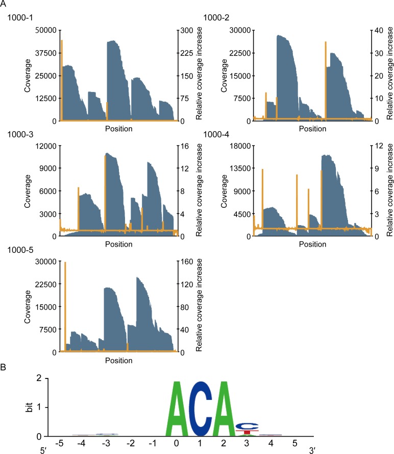
Fig 2. Identification of the MazF cleavage sequence using massive parallel sequencing.
(A) Graph of the coverage (blue bar) and the relative coverage increase (orange line). (B) Graphical representation of the conserved sequences. The nucleotide position with significant increases in coverage was numbered as zero. Twenty-five sequences were analyzed (S2 Table) and the frequency at each position was visualized with the WebLogo program [37].

