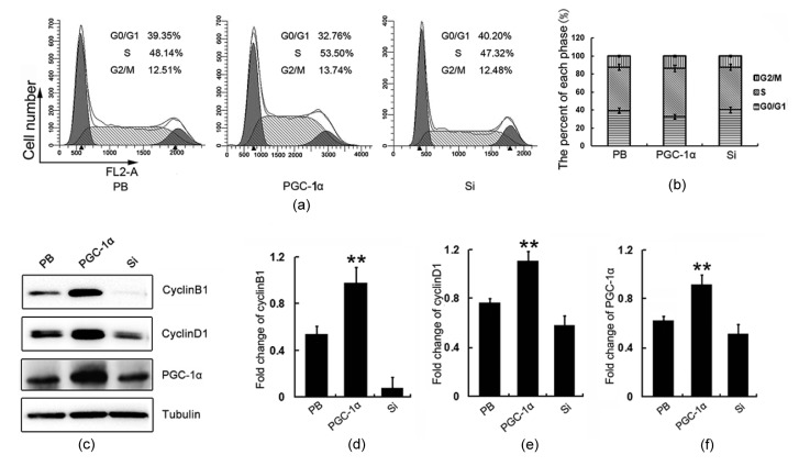
Fig. 4.
Cell cycle profiles and CylinD1/B1 expression in CH1 cells Cell cycle profiles
(a) and phase proportions (b) of PB, PGC-1α, and Si cells; (c) The expressions of CyclinD1 and CyclinB1 in PB, PGC-1α, and Si cells were detected by Western blotting; (d) Semi-quantification of CyclinB1 protein expression in (c); (e) Semi-quantification of CyclinD1 protein expression in (c); (f) Semi-quantification of PGC-1α protein expression in (c).** P<0.01. Data are expressed as mean±SD (n=3)

