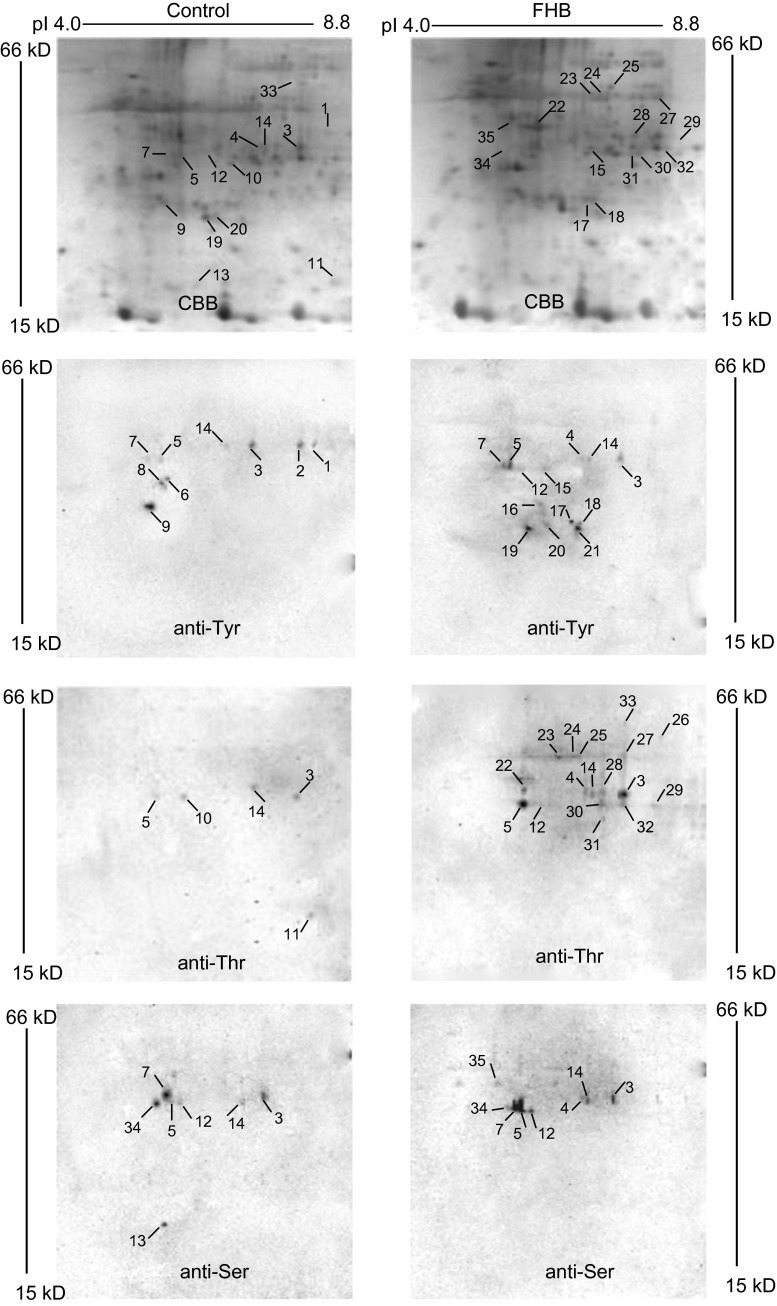
Fig. 1.
2-DE images visualized by Coomassie blue staining and immunostaining. a, b Coomassie blue-stained protein profiles of wheat spikes with water treatment and 6 hai after infection. c, d Phosphotyrosine 2-DE images of wheat spikes with water treatment and 6 hai after infection. e, f Phosphothreonine 2-DE images of wheat spikes with water treatment and 6 hai after infection. g, h Phosphoserine 2-DE images of wheat spikes with water treatment and 6 hai after infection. Representative figure from three technical and two biological replicates

