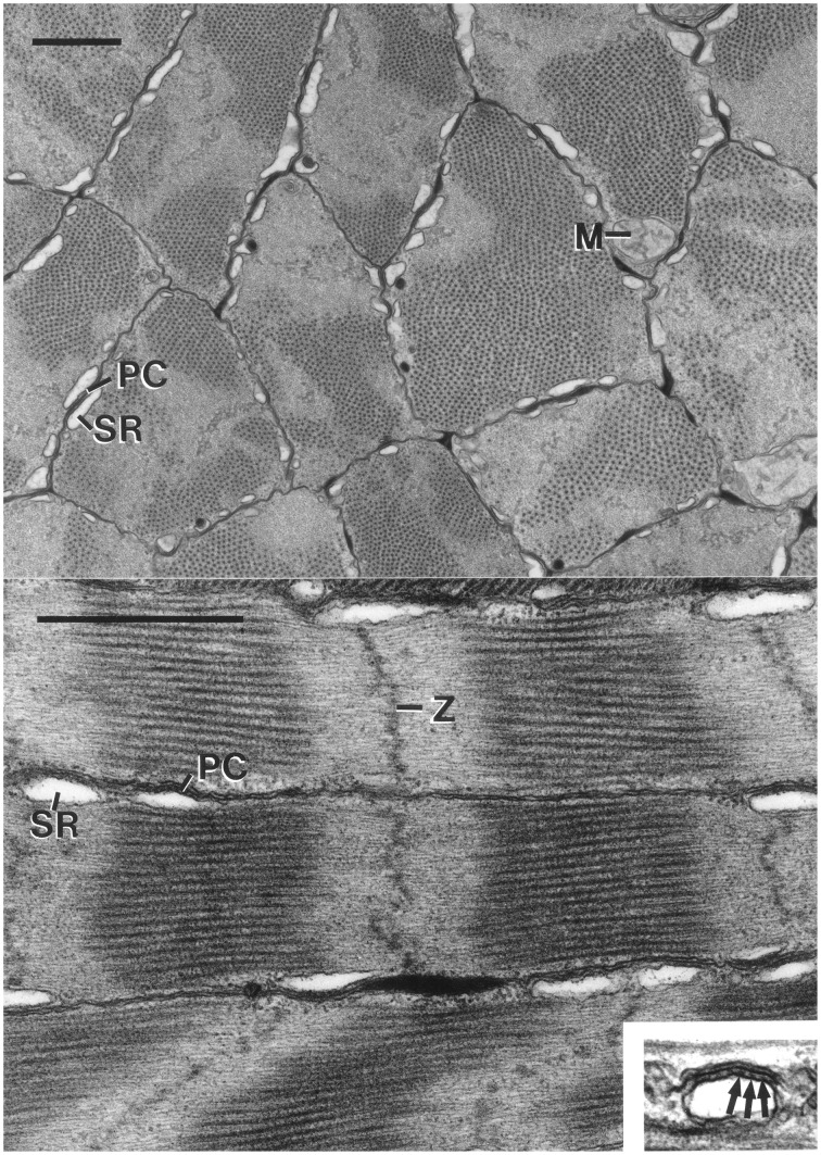
Figure 10.
Top: Electron micrograph of transverse section of the cross-striated muscle fibers of the transverse muscle mass of the tentacle of Doryteuthis pealeii. Mitochondria (M) are located immediately beneath the sarcolemma. The outer membrane of the sarcoplasmic reticulum (SR) makes specialized contacts or peripheral couplings (PC) with the sarcolemma. Note that the A band (thick filaments in cross-section) passes in and out of the section plane in a single fiber. The scale bar length equals 1 μm. Bottom: Electron micrograph of longitudinal section of cross-striated muscle fibers of the transverse musculature of the tentacle of Doryteuthis pealeii. The outer membrane of the sarcoplasmic reticulum (SR) forms peripheral couplings (PC) with the sarcolemma. The inset shows a higher magnification view of a peripheral coupling in which junctional feet (arrows) are visible. Note that the Z-disc (Z) is diffuse and sometime follows an angled course across the fiber. The scale bar length equals 1 μm and the inset is 0.5 μm wide. From Kier (1985).

