
Figure 1.
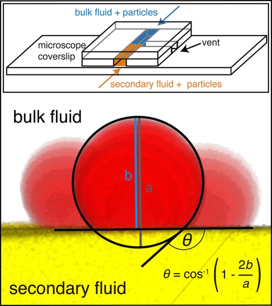
Determination of contact angle θ. The main figure shows an example 2D projection of a confocal 3D image of single particles (red) in the interface between the bulk (uncolored) and secondary (yellow) fluid. The circle and lines are added for better visualization of the geometry. The inset shows a schematic drawing of the custom-built microchannel on the microscope coverslip.